
Our Servcies
Your Journey to Health, Our Global Care: Discover seamless medical tourism with personalized services, expert consultations, and a compassionate team dedicated to your well-being.
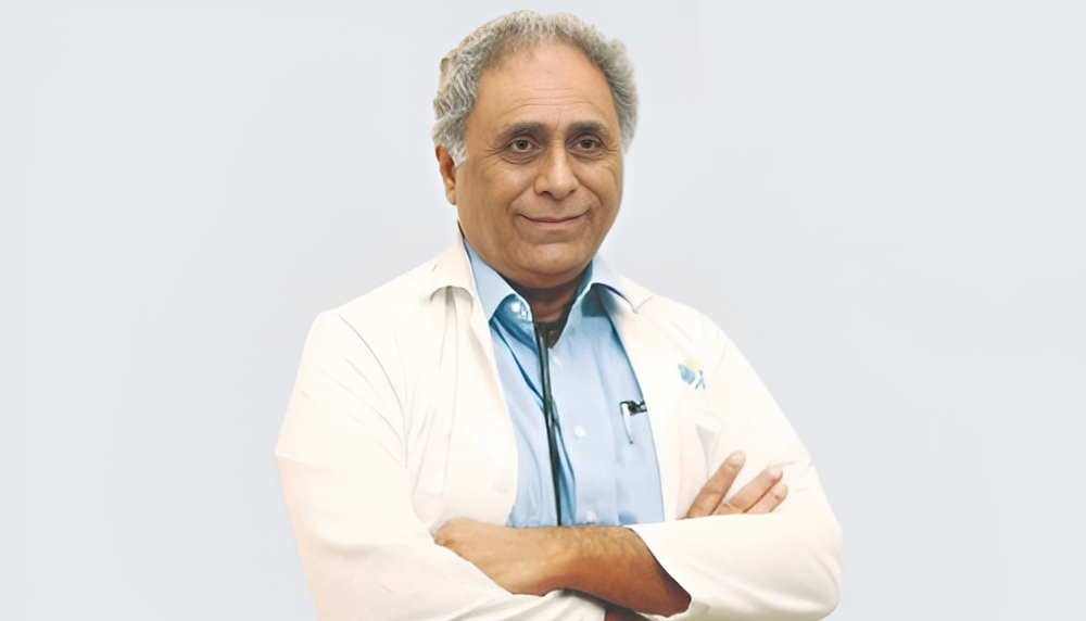
At Abrar Global Healthcare we understand the unique needs of international patients seeking medical treatment abroad. We are dedicated to providing comprehensive and personalized services to ensure a seamless and comfortable healthcare experience. From the moment you reach out to us, we offer a range of services designed to cater to your specific requirements.

Our Vision and Mission
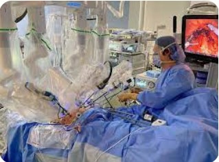
We aspire to become a global leader in medical tourism, providing exceptional healthcare access for diverse patients worldwide. Our mission is to bridge healthcare gaps, offering compassionate, world-class services and personalized care in India, catering to patients from Arabic, African, European, and Asian countries.